
- Current location:Home page >> health information
What is the difference between X-ray, CT and MRI?
2026-07-01 02:57:31
The difference between X-ray, CT and MRI: core comparison of medical imaging technology
In medical imaging technology,X-ray,CTandMRIThey are the three most commonly used inspection methods, each with its own unique advantages and applicable scenarios. X-ray is known for being fast and low-cost, and is suitable for bone and lung examination; CT provides more detailed anatomical structures through multi-layer imaging and is often used in emergency departments and tumor screening; MRI has become the first choice for nervous system and joint diseases due to its lack of radiation and high soft tissue resolution. This article will focus on the principles, indications and limitations of the three to help readers understand how to choose the most appropriate inspection method according to their needs.
X-ray: a basic but irreplaceable rapid screening tool
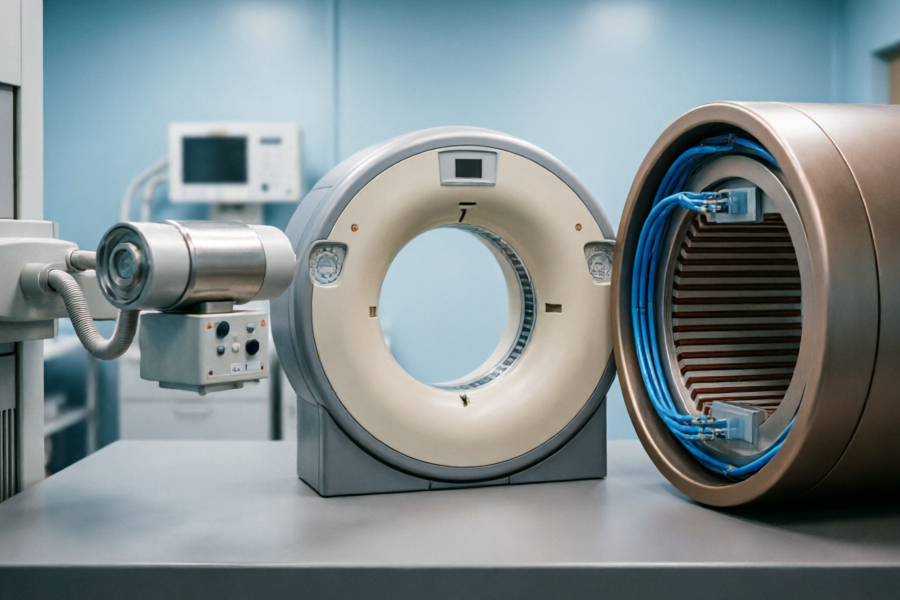
X-rays use electromagnetic waves to penetrate the human body and generate images through the differences in absorption of rays by different tissues. They mainly display denser structures such as bones and lungs. The advantage is thatfast(usually takes a few minutes to complete),low cost, suitable for fractures, pneumonia or dental check-ups. However, the limitations of X-ray are also obvious: two-dimensional images may overlap and block lesions, and the resolution of soft tissues is low. For example, early-stage lung cancer may be missed due to occlusion of the heart. At present, digital X-ray machines produced by Siemens, Philips and other manufacturers (such as SiemensMobilett Mira Max) has greatly improved imaging clarity.
CT: the “anatomy microscope” for multi-dimensional scanning
CT (computed tomography) acquires multi-angle data by rotating an X-ray source, which is reconstructed into cross-sectional images by computer, and can even generate 3D models. itshigher resolution, can clearly display blood vessels, internal organs and tiny lesions, and is often used for cerebral hemorrhage, pulmonary nodules and abdominal emergencies. For example, 64-slice CT (such as GERevolution EVO) can complete a heart scan in seconds. However, the radiation dose of CT is significantly higher than that of X-ray, so pregnant women should use it with caution. In addition, CT's soft tissue contrast performance is still inferior to MRI, and it may be difficult to identify cerebral ischemia in its early stages.
MRI: Radiation-free soft tissue 'HD camera'
MRI (magnetic resonance imaging) uses magnetic fields and radiofrequency pulses to excite hydrogen atomic signals to produce high-contrast soft tissue images, and is particularly good at examining the brain, spinal cord, joints and muscles. itsNo radiationIt is suitable for children and pregnant women and can perform multi-parameter imaging (such as T1/T2 weighting). For example, 3.0T MRI (such as SiemensMagnetom Skyra) can clearly show early cerebral infarction or ligament tear. However, MRI examinations take a long time (more than 30 minutes), require high patient cooperation, and may not be used by people with metal implants in their bodies.
Summary: Accurate selection requires weighing the pros and cons
The three technologies are complementary rather than substitutes:X-raySuitable for preliminary screening,CTBetter than fast and precise dissection,MRIWins in soft tissue and safety. The actual choice needs to take into account the condition (for example, X-ray is preferred for fractures, MRI is preferred for brain tumors), cost (X-ray is the most economical) and patient conditions (for example, MRI should be used with caution for claustrophobia). In the future, artificial intelligence-assisted diagnosis may further optimize the application efficiency of imaging technology.
Quote sources:
1. "Medical Imaging" (People's Medical Publishing House, 8th Edition)
2. American College of Radiology (ACR) Guidelines
3. Manufacturer’s product manual: Siemens Mobilet Mira Max, GE Revolution EVO, Siemens Magnetom Skyra
| technology | Principle | Advantages | limitations |
|---|---|---|---|
| X-ray | Electromagnetic wave penetration imaging | Fast and low cost | 2D overlap, low soft tissue resolution |
| CT | Multi-angle X-ray reconstruction | High-resolution, 3D imaging | Radiation is higher |
| MRI | Magnetic field excites hydrogen atom signal | No radiation, high soft tissue contrast | Long time and many contraindications |
Relevant knowledge
- 2026-10-01How do women take care of their skin scientifically?
- 2026-10-01What is female beauty?
- 2026-09-30How can women supplement calcium scientifically?
- 2026-09-30What is calcium supplementation?
- 2026-09-30How often should bone density checks be done?
- 2026-09-29How to prevent osteoporosis?
- 2026-09-29Why are women prone to osteoporosis?
- 2026-09-29What is osteoporosis?
- 2026-09-28Is hormone replacement therapy safe?
- 2026-09-28What is hormone replacement therapy?
- 2026-09-28How to manage menopause?
- 2026-09-28What are the symptoms of menopause?
- 2026-09-28What is menopause?
- 2026-09-28What does a postpartum review include?
- 2026-09-27What is a postpartum review?
- 2026-09-27Is pelvic floor muscle repair necessary?
- 2026-09-27What is postpartum pelvic floor muscle repair?
- 2026-09-27How to lose weight scientifically after giving birth?
Chinese medicinal materials
More-

View
The efficacy and function of purple beads
Overview of the efficacy and functions of purple beadsPurpurea is a plant with medicinal value. Its leaves, roots and fruits can be used as medicine. Its main functions includeStop bleeding, reduce inflammation, clear away heat and detoxify, dissipate blood stasis and reduce swellingWait.In traditional medicine, purple pearls are often used to treat symptoms such as traumatic bleeding, bruises, and sore throat. Modern research has also found that it contains active ingredients such as flavonoids
-

View
The efficacy and function of locust bean
Overview of the efficacy and functions of locust beanSophora bean, also known as Sophora japonica or Sophora japonica, is the mature fruit of Sophora japonica tree, a leguminous plant, and has various medicinal values in traditional medicine.Its main functions includeClears away heat and cools blood, moisturizes intestines and relieves constipation, lowers blood pressure, and resists oxidationetc., suitable for relieving symptoms such as hemorrhoids, bloody stools, and high blood pressure.Modern
-

View
The efficacy and function of Baiziren
Overview of the efficacy and functions of BaizirenBaiziren is the dried and mature seed kernel of Platycladus orientalis, a plant in the Cupressaceae family. Traditional Chinese medicine believes that it is flat in nature and sweet in taste, and can be used to regulate the heart, kidney, and large intestine meridians.Nourish the heart and calm the nerves, moisturize the intestines and relieve constipation, and nourish the liver and kidneysand other effects.Modern research shows that cypress seed
-
View
The efficacy and function of plum slices
Overview of the effects and functions of plum slicesPlum slices, also known as borneol or dipterocarp, is a traditional Chinese medicinal material that has the effects of rejuvenating the mind, clearing away heat and relieving pain, reducing swelling and promoting muscle growth.Its main component is dextroborneol, which is widely used in the fields of medicine, cosmetics and spices.The core functions of plum slices include relieving headaches, anti-inflammatory and sterilizing, promoting wound h
-

View
Efficacy and functions of Kochia bark
Overview of the efficacy and functions of Kochia barkKochia bark, also known as broom seedling, is a commonly used medicinal material in traditional Chinese medicine for clearing away heat and promoting dampness.Diuretic and relieve stranguria, dispel wind and relieve itching, clear away heat and detoxifyand other effects.It mainly acts on urinary system inflammation, skin itching and damp-heat jaundice, and is widely used clinically.Secondary functions include assisting in lowering blood sugar,
-

View
Efficacy and functions of Baiziren
Overview of the efficacy and functions of BaizirenBaiziren is the dried and mature seed kernel of Platycladus orientalis, a plant in the Cupressaceae family. Traditional Chinese medicine believes that it is flat in nature and sweet in taste, and can be used to regulate the heart, kidney, and large intestine meridians.Nourish the heart and calm the nerves, moisturize the intestines and relieve constipation, and nourish the liver and kidneysand other effects.Its main functions include improving sy
-

View
Eclipta Eclipta efficacy and functions
Overview of the efficacy and functions of EcliptaEclipta, also known as Eclipta or Eclipta, is a commonly used medicinal material in traditional Chinese medicine to nourish the liver and kidneys, cool blood and stop bleeding.Its core functions includeNourishes yin and nourishes kidney, blackens beard and hair, stops bleeding and reduces inflammation, the secondary effects involve improving hair loss, relieving gum bleeding and skin eczema.The primary and secondary structure is clear: liver and k
-

View
Function and role of hippocampus
Overview of the functions and functions of the hippocampusSeahorse is a precious marine creature recognized for its many benefits in both traditional medicine and modern research.Its main functions includeReplenishing kidneys and strengthening yang, anti-fatigue, regulating immunity, antioxidantetc., especially the effect of nourishing kidney and replenishing qi is the most prominent.In addition, the hippocampus also plays a certain role in improving blood circulation and relieving neurasthenia.
-
View
The effects and functions of fragrant fruit
Overview of the effects and functions of fragrant fruitXiangguo, also known as Ligusticum chuanxiong or Shannai, is a plant with both medicinal and edible value, and its rhizomes are often used in traditional Chinese medicine.Its main functions includePromotes blood circulation and removes blood stasis, dispels wind and relieves pain, improves blood circulation, the secondary effects involve relieving headaches, joint pain and women’s menstrual discomfort.The active ingredients of xiangguo, su
-

View
Efficacy and functions of Cistanche deserticola
Overview of the efficacy and functions of Cistanche deserticolaCistanche deserticola, also known as Cistanche deserticola, is a traditional Chinese medicinal material mainly produced in desert areas and is known as "desert ginseng".Its core functions includeReplenishing kidney yang, replenishing essence and blood, moistening intestines and laxative, the secondary effects involve anti-aging, regulating immunity, etc.This article will start from its medicinal value, suitable people, usage methods
-

View
White peony effect and function
Overview of the efficacy and functions of white peony rootWhite peony root is a commonly used blood-nourishing and menstrual-regulating medicine in traditional Chinese medicine.Nourishes blood and softens liver, relieves pain, astringes yin and stops sweatingIt has core functions such as irregular menstruation, hypochondriac pain, spontaneous sweating and night sweats.Its functions can be divided into two categories: primary and secondary:Main functionsIncluding regulating female endocrine, reli
-

View
Efficacy and functions of Wu Hua Xue Teng
Overview of the efficacy and functions of SpatholobusFive-flowered bloodvine is a traditional Chinese medicinal material, which mainly hasPromotes blood circulation and removes blood stasis, unblocks collaterals to relieve pain, expels wind and removes dampnessIt has other effects and is often used to treat rheumatic arthralgia, bruises and irregular menstruation.Its core function is to promote blood circulation, relieve inflammation and pain, and its secondary functions include assisting in reg