- Current location:Home page >> health information
What can an electrocardiogram detect?
2026-05-25 00:31:32
What can be found from an electrocardiogram?
Electrocardiogram (ECG) is a commonly used examination method to record the electrical activity of the heart and can help diagnose a variety of heart diseases, such as arrhythmia, myocardial ischemia, myocardial infarction, etc. The main content is divided into three categories:heart rhythm problems(such as atrial fibrillation, premature beats),Abnormal blood supply to myocardium(such as coronary heart disease) andstructural changes(eg ventricular hypertrophy). In addition, it can assist in the evaluation of electrolyte imbalances or drug effects. In primary and secondary terms, arrhythmia and ischemic changes are the core, and the others are supplementary information.
The most direct function of electrocardiogram isCatching heart rhythm abnormalities. Through waveform analysis, doctors can determine whether the heartbeat rhythm is regular. For example, atrial fibrillation shows the disappearance of P waves and unequal RR intervals; premature ventricular contractions show wide and deformed QRS waves. Holter can also record intermittent abnormalities within 24 hours, improving the detection rate of hidden problems. The American College of Cardiology points out that about 80% of paroxysmal palpitations can be diagnosed through electrocardiography.

Connecting the upper section, ECG pairMyocardial ischemia or infarctionThe diagnosis is equally critical. When coronary blood flow is blocked, ECG will show ST segment elevation or depression, T wave inversion and other characteristics. For example, "tombstone-like ST segment elevation" in acute myocardial infarction is an indication for emergency interventional treatment. According to the Lancet study, combining symptoms and ECG changes, the diagnosis rate of myocardial infarction can reach 95%. However, mild ischemia may require further confirmation with stress testing (such as exercise electrocardiography).
In addition, ECG can indirectly reflectStructural or metabolic problems with the heart. When left ventricular hypertrophy occurs, the QRS wave amplitude increases; hypokalemia manifests as an obvious U wave. Certain antiarrhythmic drugs (such as amiodarone) can prolong the QT interval and require ECG monitoring to prevent malignant arrhythmias. However, it should be noted that ECG is less sensitive to structural lesions (such as valvular disease) and is usually combined with echocardiography.
In summary, ECG is an "alarm" for heart health. Its core value lies in the rapid identification of fatal arrhythmias and ischemic events, but its judgment of the cause is limited. In clinical practice, it is often combined with other tests (such as cardiac enzymes and imaging) to improve accuracy. The CardioScript series of Bosch Medical in Germany and the ECAPS series of Nippon Optoelectronics are commonly used equipment, and their data accuracy has been certified by the FDA. As cardiologist Eugene Braunwald said: "ECG is the cornerstone of clinical decision-making, but it is not the master key."
| Common ECG Abnormalities | Corresponding disease | Typical performance |
|---|---|---|
| irregular heartbeat | Atrial fibrillation/ventricular fibrillation | Irregular RR interval |
| ST segment changes | myocardial infarction | ST elevation ≥1mm |
| Q wave broadening | old myocardial infarction | Q wave>40ms |
Quote source:
1. American College of Cardiology (ACC) "2020 Clinical Application Guidelines for Electrocardiography"
2. The Lancet "Diagnostic Criteria for Acute Coronary Syndrome" (2019)
3. Equipment manufacturers: Germany's Bosch Medical (CardioScript 12-lead meter), Nippon Kohden (ECAPS 1200)
4. Expert opinion: Eugene Braunwald's "Cardiology" 11th Edition
Relevant knowledge
- 2026-06-02What is HPV testing?
- 2026-06-02What is TCT examination?
- 2026-06-02What does a cervical smear do?
- 2026-06-01What is a cervical smear?
- 2026-06-01What is bronchoscopy?
- 2026-06-01What is capsule endoscopy?
- 2026-06-01What is painless gastroscopy?
- 2026-06-01What can a colonoscopy detect?
- 2026-05-31What is a colonoscopy?
- 2026-05-31What can a gastroscopy detect?
- 2026-05-30What is gastroscopy?
- 2026-05-30What does a bone density test mean?
- 2026-05-30What is a bone density test?
- 2026-05-30What can carotid artery color ultrasound detect?
- 2026-05-29What is carotid artery ultrasound?
- 2026-05-29What is prostate B-ultrasound?
- 2026-05-29What is gynecological B-ultrasound?
- 2026-05-29What is Breast Ultrasound?
Chinese medicinal materials
More-

View
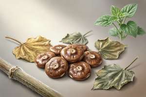
The efficacy and function of Kudidin
Overview of the efficacy and functions of KudidinKudidin is a traditional Chinese medicinal material that has the functions of clearing away heat and detoxifying, reducing swelling and relieving pain, activating blood circulation and removing blood stasis. It is often used to treat sore throat, sores, bruises and injuries. Its main active ingredients include alkaloids and flavonoids, and modern research has also found that it has antibacterial, anti-inflammatory and immunomodulatory effects. Thi
-
View
The efficacy and functions of mallow seeds
Overview of the efficacy and functions of mallow seedsMallow seeds are a traditional Chinese medicinal material whose main functions includeDiuretic and relieve stranguria, moisturize intestines and relieve constipation, clear away heat and detoxify, is commonly used in clinical Chinese medicine to treat symptoms such as difficulty urinating, edema, constipation and heat sores. Its core function comes from its rich mucus, polysaccharide and mineral components, which can gently regulate human met
-

View
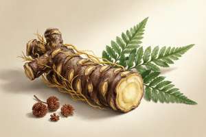
The efficacy and function of mountain turtle
### Efficacy and functions of mountain turtle #### 1. Overview Mountain turtle (scientific name: *Stephania tetrandra*), also known as "Golden Threaded Turtle" or "Fangji", is a traditional Chinese medicinal material mainly distributed in southern my country. Its roots are used as medicine and have the effects of clearing away heat and detoxifying, dispelling wind and relieving pain, diuresis and reducing swelling. It is often used to treat rheumatic arthralgia, edema, sores, swelling and poison
-
View
The efficacy and function of scalding dog spine
Overview of the efficacy and functions of hot dog spineHot dog spine is a processed product of the traditional Chinese medicine dog spine. It hasNourish liver and kidney, strengthen muscles and bones, remove rheumatismWith core functions such as this, it is often used to treat related symptoms caused by soreness and weakness in the waist and knees, rheumatic arthralgia and kidney deficiency. Its mechanism of action is mainly due to the active ingredients such as flavonoids and volatile oils, whi
-
View
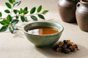
Effects and functions of vinegar and myrrh
Below is structured content about the benefits and effects of vinegar and myrrh, generated as per your request:First paragraph: content overviewVinegar myrrh is a commonly used processed product in traditional Chinese medicine. It is processed from myrrh through vinegar. Its main functions are to promote blood circulation and relieve pain, reduce swelling and promote muscle growth. Its core functions are reflected in three aspects: first, it relieves blood stasis pain by promoting blood circulat
-
View
The efficacy and function of Daochi Powder
Overview of the efficacy and functions of Daochi PowderDaochi Powder is one of the classic prescriptions of traditional Chinese medicine. It is mainly used to clear away heat and dampness, relieve stranguria and relieve pain. It is especially good at treating symptoms such as short red urine, sores on the mouth and tongue caused by excessive heart fire or damp heat. Its core functions includeClear the heart, reduce fire, diuretic and relieve stranguria, its secondary function is to relieve urina
-

View
The efficacy and function of dream flower
The efficacy and function of dream flower: the multiple values of traditional herbal medicineDream flower, also known as knotweed or yellow daphne, is a plant with both ornamental and medicinal value. Its core functions includeRelaxing muscles and activating collaterals, reducing swelling and relieving pain, calming nerves and promoting sleep, secondary effects involve antibacterial, anti-inflammatory and improved skin health. The primary and secondary structure is clear: medicinal value is the
-

View
The efficacy and functions of Huangjingzi
Overview of the efficacy and functions of HuangjingziHuang Jing Zi, also known as Bu Jing Zi, is the dried and mature fruit of Vitex japonicus, a plant of the Verbenaceae family, and is widely used in traditional Chinese medicine. Its nature and flavor are pungent, bitter, and warm, and returns to the lung, stomach, and liver meridians.Expelling wind and relieving surface, resolving phlegm and relieving cough, promoting qi and relieving pain, resolving dampness and neutralizingIt has other effec
-
View
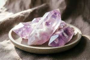
The efficacy and function of amethyst
Overview of the efficacy and functions of amethystAmethyst is a traditional Chinese medicine with a long history of medicinal use. Its main functions includeCalming the mind and calming the nerves, warming the lungs and relieving asthma, warming the palace and dispersing cold, suitable for palpitations, insomnia, cough and asthma due to deficiency and cold, and uterine cold in women. Its core role stems from the containedcalcium fluorideAnd other mineral components, modern research has also foun
-

View
The efficacy and role of artemisinin
The efficacy and role of artemisinin: from anti-malaria to the exploration of modern medicineArtemisinin is an active ingredient extracted from the traditional Chinese medicine Artemisia annua. Its most significant effect isAnti malaria, especially has a significant effect on drug-resistant malaria parasites. In addition, research has also found that it has certain value in the fields of anti-inflammation, immune regulation and potential anti-tumor. This article will start with its core function
-

View
Effects and functions of sarsaparilla
Overview of the efficacy and functions of sarsaparillaSarsaparilla is a traditional Chinese medicinal material that has the effects of dispelling wind, removing dampness, reducing swelling and relieving pain, and benefiting joints. It is often used as an auxiliary treatment for rheumatism, bruises, and urinary system diseases. Its main active ingredients include flavonoids, saponins, etc. Modern research has also found its antioxidant and anti-inflammatory effects. In terms of content structure,
-
View
The efficacy and function of fried Citrus aurantium husk
Overview of the efficacy and functions of fried Fructus AurantiiStir-fried Citrus aurantium is a qi-regulating medicine commonly used in traditional Chinese medicine. It is made from Citrus aurantium and has the functions ofExpands the circulation of qi, relieves swelling and fullness, resolves phlegm and relieves stagnationand other core functions. It is mainly used for symptoms such as stagnation of spleen and stomach qi, fullness of the chest and hypochondrium, and indigestion. It is especial